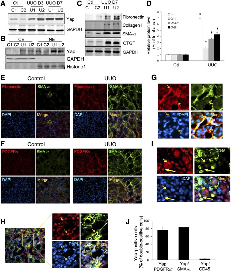
Figure 1.
Activated Yap colocalizes with interstitial cells in UUO kidneys. (A) UUO stimulates Yap expression. (B) UUO induces Yap nuclear translocation. After 7 days of UUO, the cytoplasmic and nuclear proteins were prepared and the levels of Yap in kidneys were detected by western blot analysis. GAPDH and histone 1 were used as loading controls for cytoplasm and nuclear, respectively. (C and D) UUO stimulates myofibroblast accumulation and ECM protein deposition. The myofibroblast marker (SMA-α) and ECM proteins were determined by western blot (C) with density analysis shown (D). (E and F) Double immunofluorescent staining of SMA-α with fibronectin (E) or PDGFRα (F) in control and obstructed kidneys after 7 days of UUO. (G and H) Double immunofluorescent staining of Yap in obstructed kidneys with myofibroblast marker SMA-α (G), PDGFRα (H), and inflammatory monocyte marker CD45 (I), respectively (arrowhead indicates Yap staining in interstitial cells and tubules). (J) The percentage of Yap-positive cells in the indicated double-positive cells was counted and calculated. *P<0.05 versus control; n=5. CE, cytoplasmic extract; NE, nuclear extract.

