Figure 2.
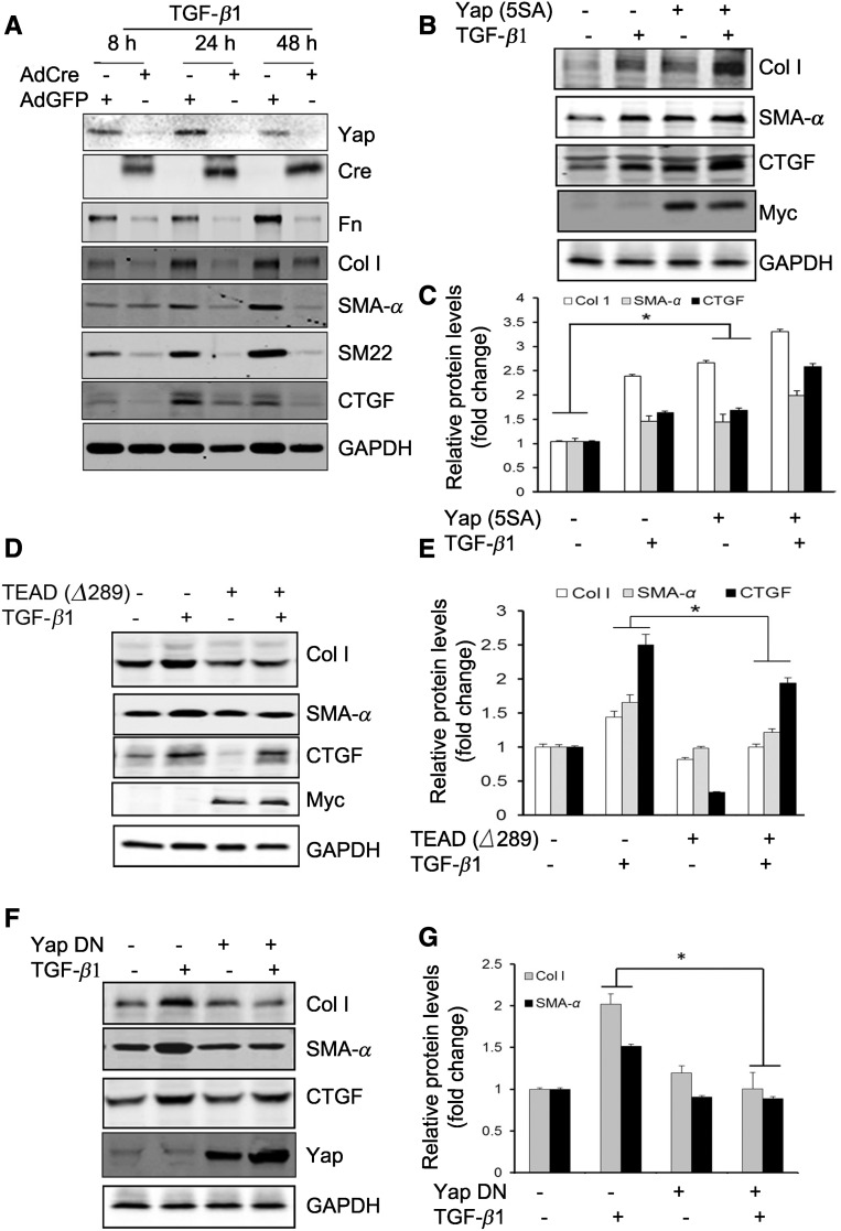
Yap mediates TGF-β1–induced fibroblast activation. (A) Mouse fibroblasts were isolated from Yapf/f/Tazf/f mice. The cells were infected with Adeno-Cre; the Adeno-GFP was used as control. After 48 hours, the cells were serum starved and treated with TGF-β1 (2 ng/ml), cell lysates were collected at the indicated time points, and the levels of indicated molecules were determined by western blots. (B and C) Mouse fibroblasts expressing constitutive active Yap (5SA) promoted fibroblast transformation into myofibroblasts. Density analysis of the western blots is shown (C). (D and E) Mouse fibroblasts overexpressing dominant negative TEAD (Δ289) suppressed the expression of collagen I, SMA-α, and Yap target CTGF (D). The corresponding density analyses of the western blots are shown (E). *P<0.05 versus TGF-β1–treated group; n=3 repeats. (F) Overexpression of dominant negative Yap (Δ60–89) suppressed TGF-β1–induced myofibroblast activation. The density analysis of the levels of collagen I and SMA-α are shown in (G). *P<0.05; n=3 repeats. AdCre, adenovirus overexpressing Cre; AdGFP, adenovirus overexpressing GFP; DN, dominant negative.

