Figure 1.
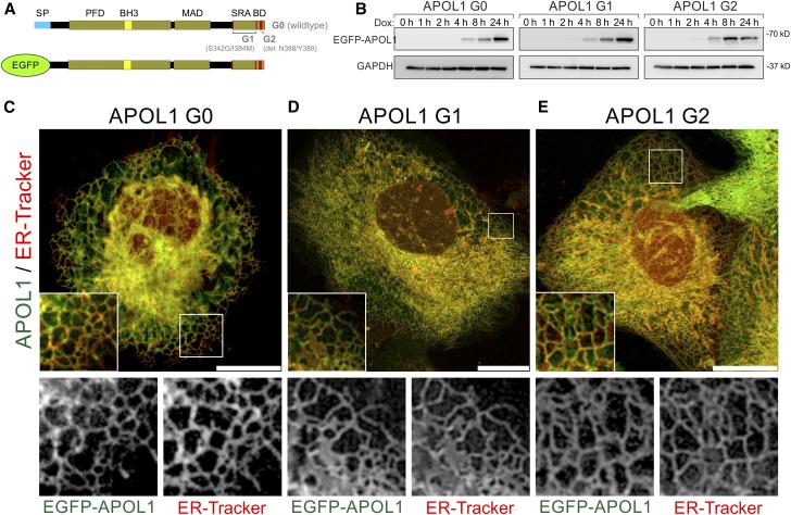
EGFP-tagged APOL1 lacking the SP is targeted to the ER in podocytes. (A) Scheme of EGFP APOL1 fusion proteins used, in which the endogenous N-terminal SP (blue) was replaced by EGFP (green). All variants include the PFD and MAD. APOL1 RRV–associated G1 point (S342G/I384M) and G2 deletion (Δ388N389Y) mutants are localized in the C-terminal SRA-BD. (B) Western blot analyses demonstrated that expression of APOL1 G0 and RRVs G1 and G2 starts 4 hours after adding doxycycline to the cell culture medium. Expression reached a maximum after 24 hours. (C–E) Live cell image analyses of EGFP-tagged APOL1 (green, Dox induction 24 hours) in combination with ER-Tracker (red) revealed a predominant localization of APOL1 to tubular-like and perinuclear localized membrane structures (C–E, details) of the ER. Noncytotoxic EGFP-APOL1 G0 and RRVs APOL1 G1 (D) and G2 (E) showed a similar intracellular distribution. Scale bar and square length of details are 20 and 10 µm, respectively.

