Figure 4.
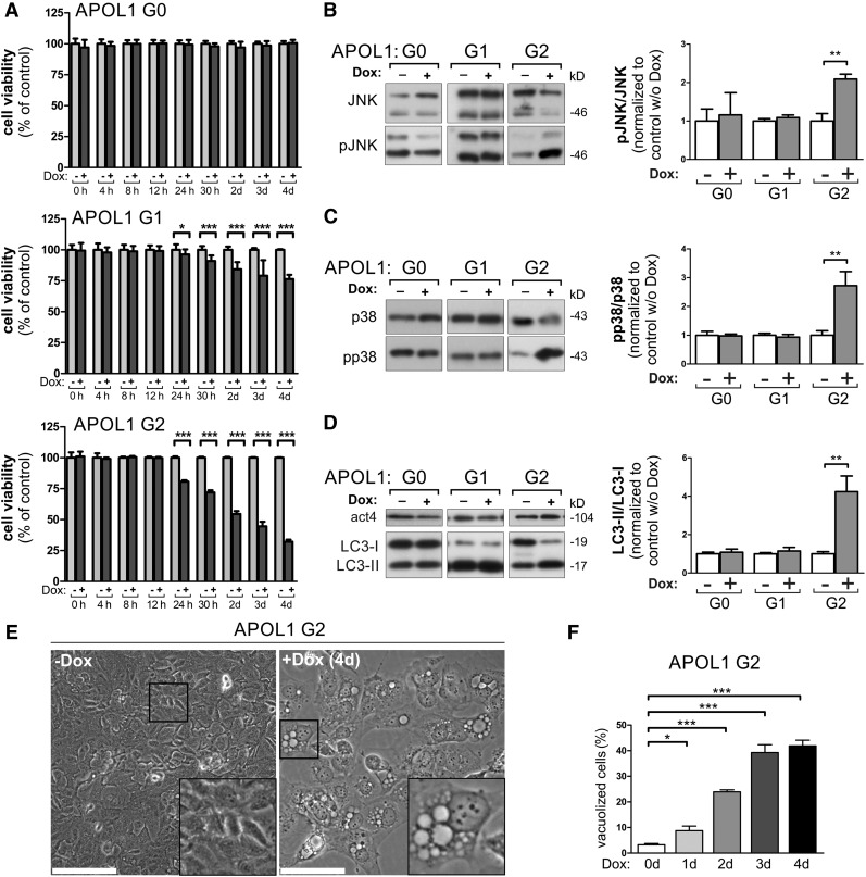
APOL1 RRVs lacking the SP cause reduced cell viability in podocytes. (A) Cytotoxicity analyses with stable podocyte cell lines, enabling the doxycycline-dependent overexpression of EGFP-APOL1 G0 and RRVs, revealed that APOL1 RRVs were able to cause a significant reduction of the cell viability. Podocytes were less susceptible to an APOL1-linked reduced viability than HEK293T cells (see Figure 3A), because the effect is not detectable in G0-overexpressing cells. Thus, AB8 podocytes were more resistant against APOL1-linked cytotoxic effects. In all experiments, the effect was stronger in APOL1 G2–overexpressing cells. (B–D) Western blot analysis (left) and quantifications (right). Lysates from EGFP-APOL1–overexpressing podocyte (Dox induction 24 hours) cells elucidated an elevated phosphorylation of stress kinases pJNK/pSAPK (B), and p-p38 (C), and an accumulation of the autophagy marker protein LC3-II (increased LC3-II/LC3-I ratio) (D) in cells overexpressing APOL1 G2. (E) Transmitted light bright-field images of podocytes expressing APOL1 G2 for 4 days show a strong vacuolization phenotype. Scale bars represent 100 µm. (F) Quantification over 4 days of doxycycline induction indicates progressive cellular vacuolization induced by APOL1 G2 in podocytes. Data are shown as mean±SEM of at least three independent experiments; *P<0.05, **P<0.01, ***P<0.005. w/o, without.

