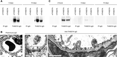
Figure 4.
Antibody elution and ultrastructural changes after exposure to anti-THSD7A IgG. (A) Western blot analysis of reactivity of IgG eluted from frozen kidney sections 5 and 14 days after injection of preimmune or anti-THSD7A IgG (PI IgG and THSD7A IgG, respectively) with recombinant mTHSD7A and hTHSD7A. (B) Electron microscopic studies of mice 2 weeks after injection of preimmune or anti-THSD7A IgG. Arrows in the lower left panel indicate subepithelial electron-dense deposits. Scale bars indicate 1 µm. (C) Western blot analysis of reactivity of mouse sera with mTHSD7A and hTHSD7A 2 hours, 5 days, and 14 days after exposure to PI IgG and THSD7A IgG. An HRP-conjugated anti-rabbit IgG antibody was used as the secondary antibody to detect circulating rabbit anti-THSD7A IgG.

