Figure 5.
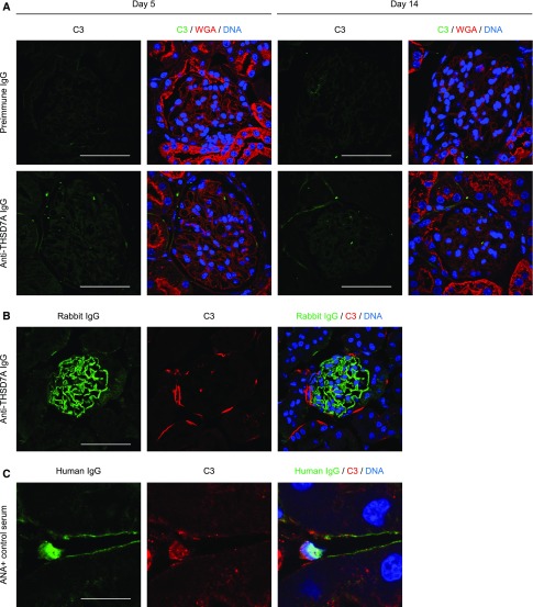
Anti-THSD7A antibodies and complement activation. (A) Immunofluorescence staining for C3, wheat germ agglutinin (WGA), and DNA in mice 5 days and 14 days after injection of preimmune IgG or anti-THSD7A IgG (C3 [FITC], WGA [Rhodamine], DNA [Draq5]). Scale bars indicate 50 µm. (B) Immunofluorescence staining for rabbit IgG, C3, and DNA after incubation of cryosections from naïve murine kidneys with anti-THSD7A IgG and complement-containing full human serum (Rabbit IgG [AF488], C3 [CY3], DNA [Draq5]). Scale bars indicate 50 µm. (C) Immunofluorescence staining for human IgG, C3, and DNA after incubation of cryosections from naïve murine kidneys with ANA-containing human serum and complement-containing full human serum (Human IgG [CY2], C3 [CY3], DNA [Draq5]). Scale bars indicate 12.5 µm.

