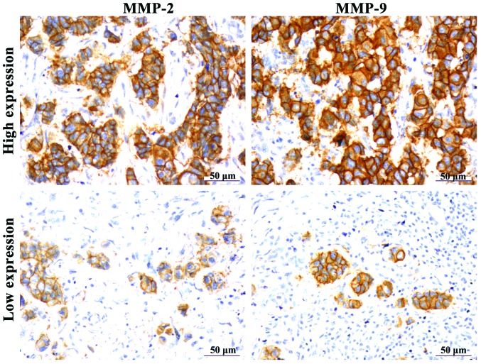
Figure 3.
Detection of matrix metalloproteinase-2 (MMP-2) and MMP-9 protein expression in clinicopathological tissues via immunohistochemistry (IHC) MMP-2 and MMP-9 are expressed in the cytoplasm and appear as brown granules; a proportion of positive cells >25% in the visual field represented high expression, otherwise, the result was categorized as low expression (×400).

