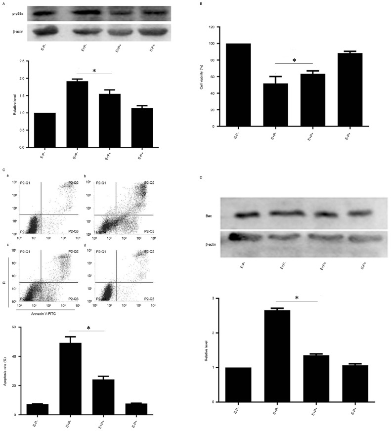
Figure 3.
Effects of p38α inhibition on EGCG-mediated apoptosis and protein expression levels of Bax and p-p38α in NB4 cells. NB4 cells were pretreated with 10 µM PD169316 for 0.5 h and then treated with 30 µM EGCG for 24 h. (A and D) Western blot analysis was used to detect the protein expression level of p-p38α and Bax. (B) Cell-Counting Kit-8 assay was used to measure the cell viability of NB4 cells. (C) Flow cytometric analysis was used to determine apoptotic rate in NB4 cells. a, EGCG− PD169316−; b, EGCG+ PD169316−; c, EGCG+ PD169316+; d, EGCG− PD169316+. All experiments were performed in triplicate. *P<0.05 vs. control group. EGCG, epigallocatechin-3-gallate; Bax, Bcl-2-like protein 4; E, 30 µM EGCG. P, 10 µM PD169316; PI, propidium iodide; FITC, fluorescein isothiocyanate.

