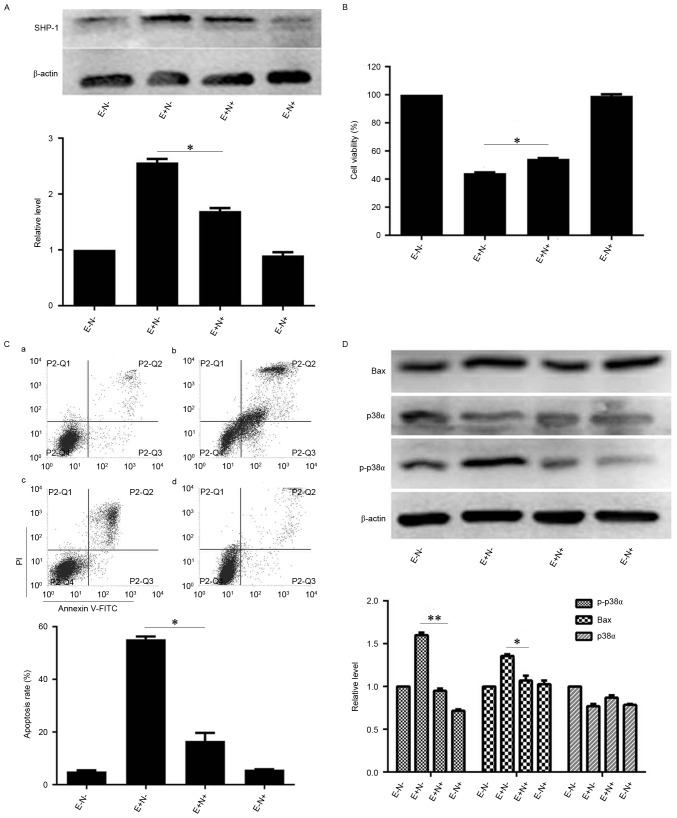
Figure 4.
Effects of SHP-1 inhibition on EGCG-mediated apoptosis and expression levels of associated proteins in NB4 cells. NB4 cells were pretreated with 10 µM NSC87877 for 0.5 h and then treated with 30 µM EGCG for 24 h. (A) Western blot analysis was used to detect the expression level of SHP-1. (B) Cell Counting Kit-8 assay was used to measure the viability of NB4 cells. (C) Flow cytometric analysis was used to determine the apoptotic rate of NB4 cells. a, EGCG− NSC87877−; b, EGCG+ NSC87877−; c, EGCG+ NSC87877+; d, EGCG− NSC87877+. (D) p38α, p-p38α and Bax protein levels were assessed by western blot analysis. All experiments were performed in triplicate. *P<0.05, **P<0.01 vs. control group. EGCG, epigallocatechin-3-gallate; SHP-1, Src homology region 2 domain-containing phosphatase-1; p38α, p38α mitogen activated protein kinase; E, 30 µM EGCG. N, 10 µM NSC87877; p-, phosphorylated.

