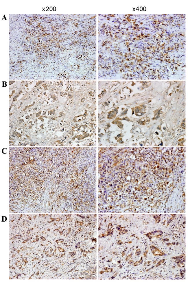
Figure 3.

Immunohistochemical staining for HOXA9 and PBX3 in GC and in normal tissue. (A) Immunostaining of HOXA9 in poorly-differentiated GC tissue revealed that positive staining was mainly in the nucleus and also in the cytoplasm. (B) Immunostaining of HOXA9 in moderately-differentiated GC tissue. Positive staining was mainly in the nucleus and cytoplasm. (C) Immunostaining of PBX3 in poorly-differentiated GC tissue. Positive staining was mainly in the nucleus and also in the cytoplasm. (D) Immunostaining of PBX3 in moderately differentiated GC tissue and positive staining was mainly in the nucleus and cytoplasm. GC, gastric cancer; HOXA9, homeobox A9; PBX3, PBX homeobox 3. Magnification, ×200 (left) and ×400 (right).
