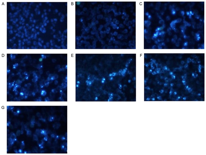
Figure 4.
Effect of shRNA treatment on morphological changes of apoptotic cells by Hochest 33258 stain analysis. (A) blank control; (B) shNC; (C) shRNA2; (D) shRNA3; (E) shRNA4; (F) shRNA5; and (G) shRNA6. The cells treated with shRNAs exhibited changes typical of apoptotic cells following transfection: Cell shrinkage, nuclear condensation and dense stain with some white color. The fluorescence staining of the shNC and blank control groups was light and uniform.

