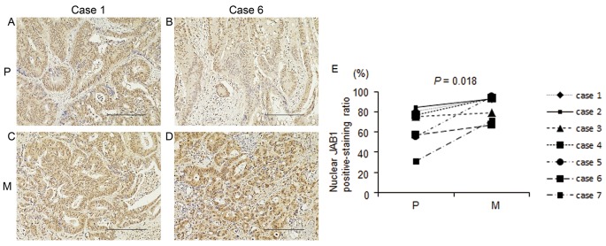
Figure 4.
Immunohistochemical analysis of JAB1 expression in primary colorectal cancer tissues and liver metastases following mainly 5-FU-based chemotherapy. (A-D) Representative images of JAB1 staining in primary colorectal cancer tissues (A and B) and in liver metastases (C and D). Primary colorectal cancer tissue (A) and liver metastasis (C) are derived from the same patient (case 1). Primary colorectal cancer tissue (B) and liver metastasis (D) are also paired (case 6). Magnification, ×40. Scale bar, 200 µm. (E) The proportion of cells positive for nuclear JAB1 expression in primary colorectal cancer tissues and liver metastases following mainly 5-FU-based chemotherapy (n=7). P, primary colorectal cancer tissue; M, liver metastasis. JAB1, Jun activation domain-binding protein 1; 5-FU, 5-fluorouracil.

