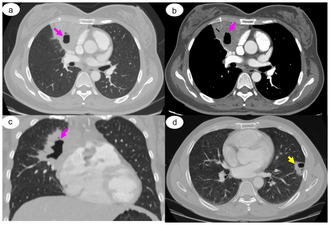
Figure 3.
Representative CT images. Case of a 45-year-old woman with primary mucosa-associated lymphoid tissue lymphoma, confirmed by CT-guided transthoracic needle biopsy. Chest CT revealed a well-defined lesion with consolidation and cavitation in the right upper lobe of the lung (arrow) in (A) pulmonary window, (B) soft-tissue window and (C) coronal pulmonary window. (D) Case of a 43-year-old man with primary pulmonary Hodgkin's lymphoma, nodular sclerosis type, confirmed by bronchoscope biopsy. Chest CT revealed a peripherally located 4.5-cm mass with cavitation in the left lower lobe of the lung (arrow).

