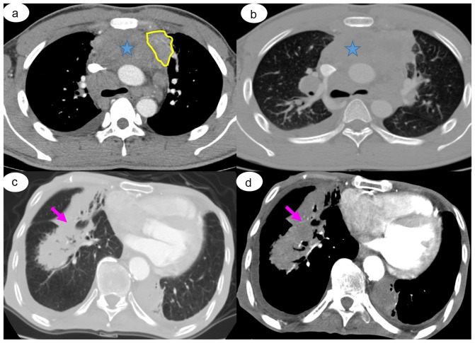
Figure 4.
Representative CT images. Case of a 47-year-old man with secondary pulmonary diffuse large B lymphoma derived from the right axillary lymph nodes, confirmed by bronchoscopic biopsy. (A) Chest CT images revealed an ill-defined mass on the upper lobe of the left lung (yellow area) with multiple mediastinal lymphadenopathy. (B) Fusion and necrosis of the enlarged mediastinal lymph nodes (blue star) and a small amount of pleural effusion posteriorly was observed. Case of a 56-year-old man with secondary pulmonary non-Hodgkin's lymphoma, confirmed by bronchoscopic biopsy. Chest CT revealed bilateral diffuse lesions with irregular edges and an air bronchogram (arrow) in (C) pulmonary window and (D) soft-tissue window.

