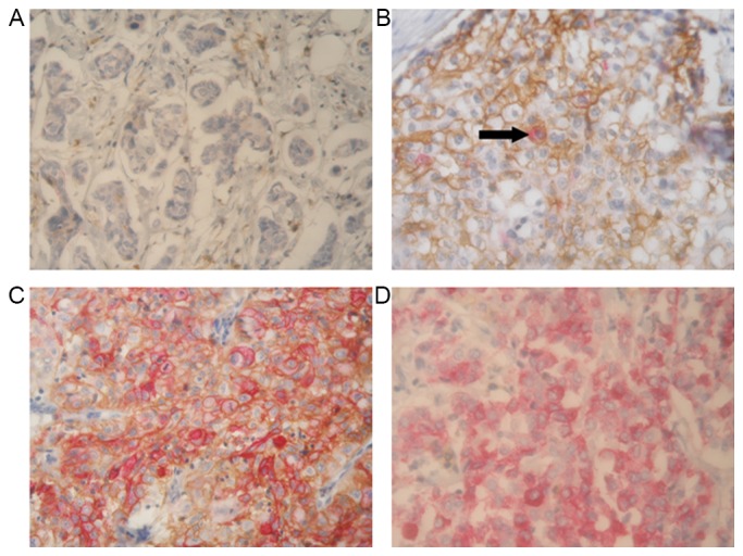
Figure 2.

Representative immunohistochemical double-staining patterns of four CD44/CD24 phenotypes in triple-negative breast cancer tissues. CD44 (brown) exhibited homogenous membranous distribution, and CD24 (red) showed membranous and cytoplasmic immunoreactivity. (A) A tumor representing the CD44−/CD24− phenotype. (B) The predominant cells are CD44+/CD24− cells, only a few cells are CD44+/CD24+ (black arrow). (C) A tumor tissue considered as representing the CD44+/CD24+ phenotype. (D) Almost all cells in this tumor are CD44−/CD24+ cells. Magnification, ×400. CD, cluster of differentiation.
