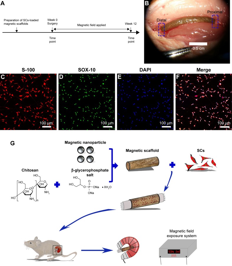
Figure 1.
Schematic of this study.
Notes: (A) Time scale. (B) The SCs-loaded magnetic scaffold bridging 15 mm sciatic nerve defect in rats under the microscope. (C–E) Double immunofluorescent staining shows positive S-100 and SOX-10 with DAPI nuclear counterstaining. (F) Merged image shows a high purity of SCs (>97%). (G) The schematic diagram of the main process of the experiment.
Abbreviations: DAPI, 4′,6-diamidino-2-phenylindole; SCs, Schwann cells.

