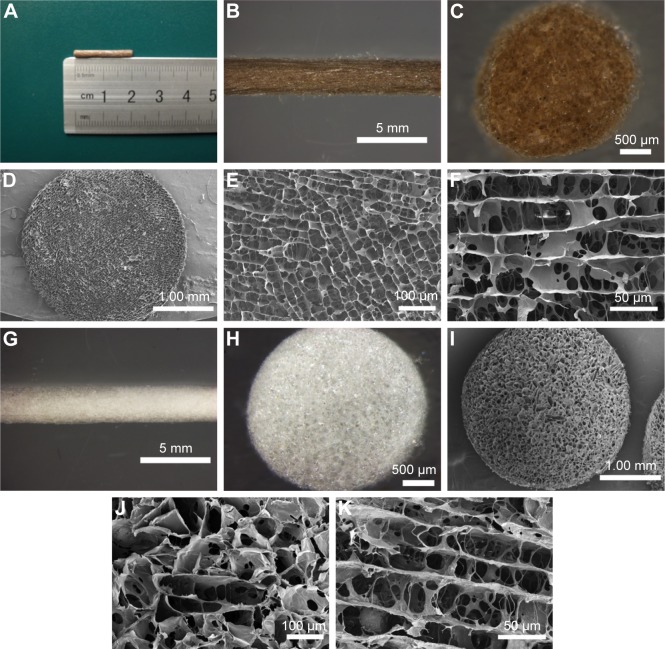
Figure 3.
The morphology of the nonmagnetic and magnetic scaffolds.
Notes: (A) The length of the magnetic scaffold. (B, C, G, and H) The general and cross-sectional appearance of the magnetic and nonmagnetic scaffolds under stereomicroscope, respectively. (D and I) The general structures of the magnetic and nonmagnetic scaffolds under SEM, respectively. (E and J) Representative transverse photographs of the magnetic and nonmagnetic scaffolds, showing that the microarrays were formed in a honeycomb-shaped characteristic. (F and K) Representative longitudinal photographs of the magnetic and nonmagnetic scaffolds, displaying the interconnected cellular architecture and lengthwise oriented microchannels.
Abbreviation: SEM, scanning electron microscopy.

