Figure 1.
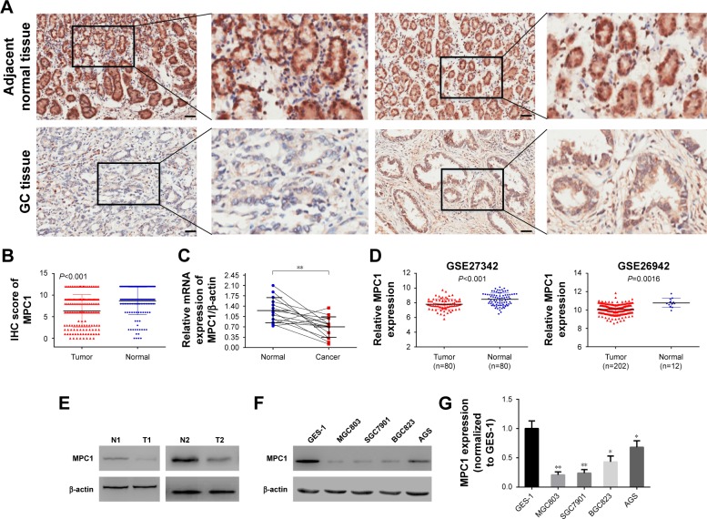
The protein and mRNA levels of MPC1 were significantly decreased in GC tissues and cell lines.
Notes: (A) Representative images of immunohistochemistry (IHC) staining of MPC1 in GC and adjacent normal tissues. Scale bar, 50 μm. (B) IHC scores of MPC1 of adjacent normal tissues and cancerous tissues in 152 paired GC specimens (P<0.001). (C) MPC1 mRNA levels in 15 pairs of fresh GC specimens and adjacent normal tissues (P<0.01). (D) Analyses of MPC1 mRNA levels in GSE27342 and GSE26942 (P<0.001, P=0.0016, respectively). (E) Western blotting analysis of MPC1 in two pairs of GC and adjacent normal tissues. (F) Detection of MPC1 in GC cell lines (SGC7901, MGC803, BGC823, and AGS) and gastric epithelium cell line (GES-1) by Western blotting analysis. (G) Detection of MPC1 in GC cell lines (SGC7901, MGC803, BGC823, and AGS) and GES-1 by qRT-PCR analysis. *P<0.05, **P<0.01.
Abbreviations: GC, gastric cancer; IHC, immunohistochemistry.

