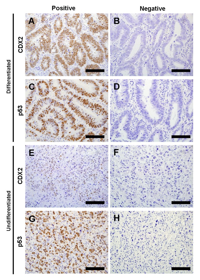
Figure 1.
Representative images of immunohistochemical staining of CDX2 and p53 in gastric cancer tissues. (A) Positive and (B) negative CDX2 staining in differentiated gastric cancer tissues. (C) Positive and (D) negative p53 staining in differentiated gastric cancer tissues. (E) Positive and (F) negative CDX2 staining in undifferentiated gastric cancer tissues. (G) Positive and (H) negative p53 staining in undifferentiated gastric cancer tissues. Scale bar, 100 µm. CDX2, caudal-type homeobox protein 2.

