Figure 4.
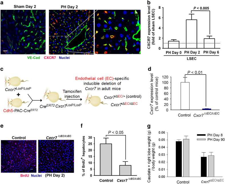
After PH, SDF-1 receptor CXCR7 is upregulated in LSEC and contributes to hepatocyte proliferation. (a) Two days after PH, the liver sections were stained for endothelial-specific VE-cadherin (green fluorescence). VE-cadherin+ LSECs are co-localized with CXCR7+ LSECs (red fluorescence). Scale bar, 50 μm. (b) Cxcr7 mRNA level in isolated LSECs was examined at indicated time after PH. The Cxcr7 expression level of sham LSEC was arbitrarily defined as 1. N=5–7 mice per group. (c, d) Mice harboring loxP-flanked Cxcr7 were crossed with endothelial cell-specific Cdh5-PAC-CreERT2 mice.58 Generated offsprings were treated six times with tamoxifen injection (250 mg kg−1) to induce Cxcr7 deletion (Cxcr7iΔEC/iΔEC).15 Mice carrying endothelial haplodeficiency of Cxcr7 (Cxcr7iΔEC/+) were used as control group. N=4 mice per group. (e–g) Inducible knockout of Cxcr7 in LSEC abrogated hepatocyte proliferation (e, f) and restoration of liver mass at indicated time after PH (g). Prohibition of liver mass recovery in Cxcr7iΔEC/iΔEC mice persisted for up to 90 days after PH; N=6–8 mice per group, P<0.01 between control and Cxcr7iΔEC/iΔEC group.

