Figure 1.
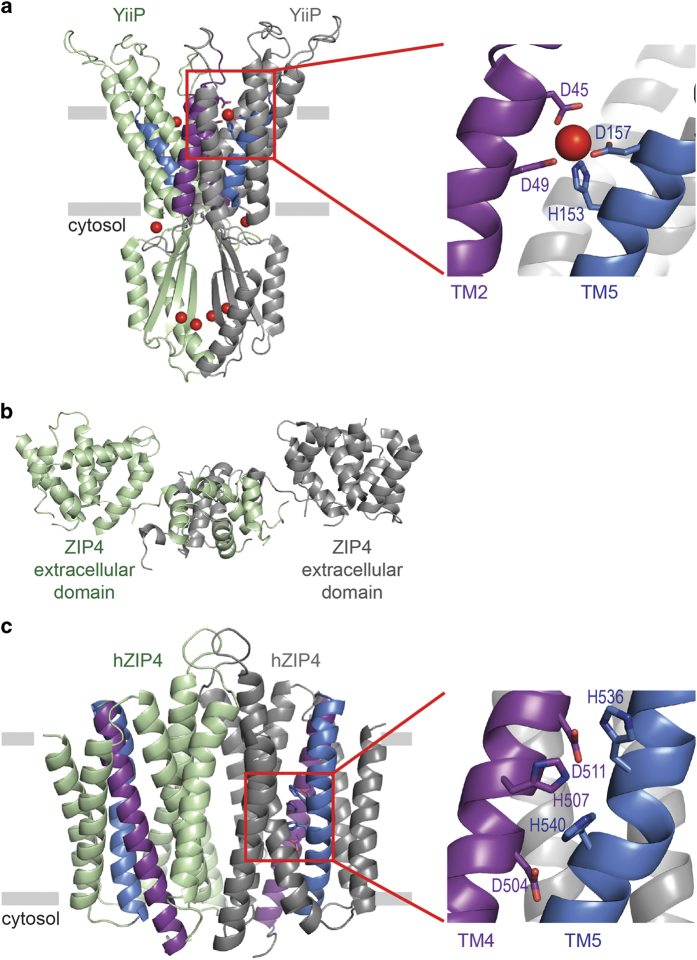
Structural basis for zinc transport by ZnT and ZIP proteins. (a) Crystal structure of bacterial ZnT homolog showing zinc (red spheres) bound in the intracellular domain, the membrane interface and TM domain. TM2 (purple) and TM5 (blue) are involved in zinc ion coordination.78 (b) Crystal structure of mammalian ZIP4 N-terminal ectodomain.81 (c) Structural model of human ZIP4 TM domain.80 TMs 2, 4, 5 and 7 are located in the center of each monomer, and TM4 (purple) and TM5 (blue) contain conserved histidine and aspartic acid residues predicted to coordinate zinc.

