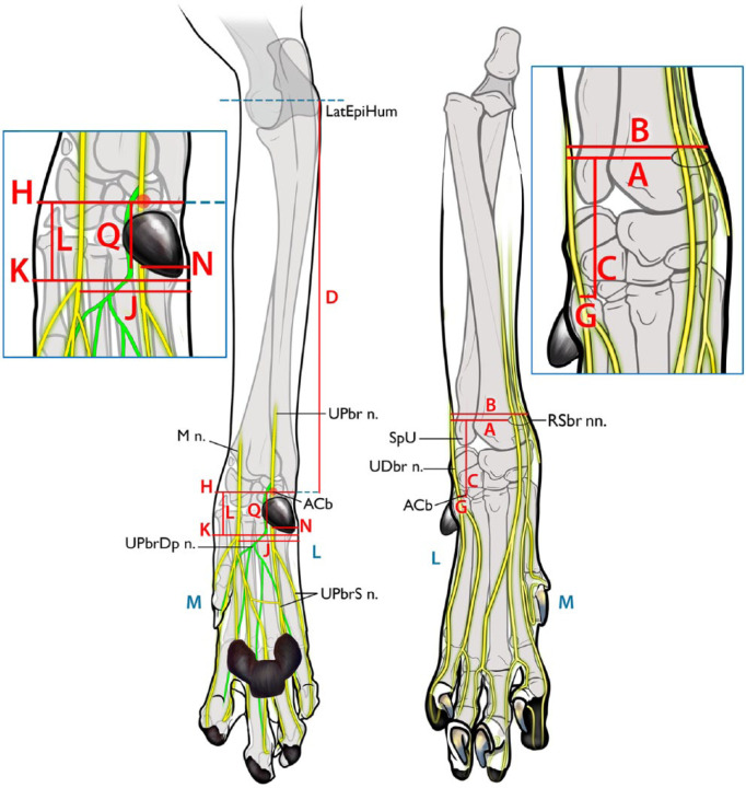
Figure 1.
Description of the measured parameters used to evaluate the proposed injection sites for distal limb nerves. (Right) Craniocaudal view. (A) Distance from the lateral aspect of the limb to the superficial branches of the radial nerve (RSbr nn.); (B) width of the limb at the level of the injection point of the RSbr nn.; (C) Distance proximally from the accessory carpal bone (ACb) to the RSbr nn.; (G) distance from the accessory carpal bone to the dorsal branch of the ulnar nerve (UDbr n.). (Left) Caudocranial view. (D) distance from the accessory carpal bone to the lateral epicondyle of the humerus (LatEpiHum); (H) width of the carpus at the level of the accessory carpal bone; (J) distance from the lateral aspect of the limb to the median nerve (M n.); (K) width of the limb at the level of the injection point of the M n. and the superficial branch of palmar branch of ulnar nerve (UPbrS n.); (L) proximodistal distance from the accessory carpal bone to the level of the M n. and UPbrS n.; (N) distance from the lateral aspect of the limb to the UPbrS n.; (Q) distance from the accessory carpal bone to the point where the deep branch of palmar branch of ulnar nerve (UPbrDp n.) turns to a deeper location. L (blue) = lateral; M (blue) = medial; SpU = styloid process of the ulna; UPbr n. = palmar branch of the ulnar nerve

