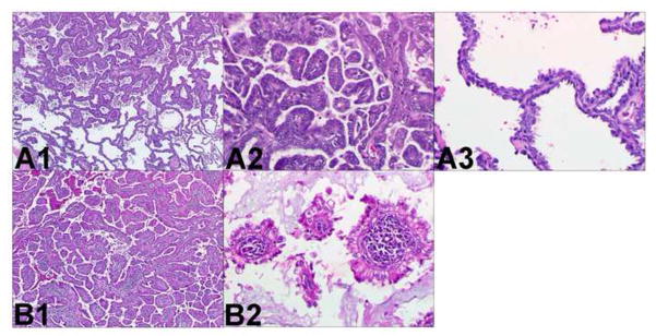
Figure 2.
Two different adenocarcinomas. Comprehensive histologic review of case 1 (paraffin-embedded, formalin-fixed tissues, hematoxylin and eosin staining). The following histologic subtypes of adenocarcinoma are identified in tumor A (panel A1, x40): 60% papillary (panel A2, x200) and 40% bronchioloalveolar (panel A3, x200). The following histologic subtypes are identified in tumor B (panel B1, x40): 80% papillary (panel B2, x200), 10% acinar, and 10% bronchioloalveolar. In addition there is prominent lymphoid stroma. These different morphological features suggest these tumors are multiple primary lung cancer.

