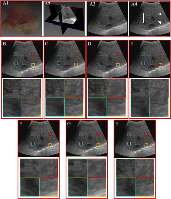
Fig. 4.

Comparison of different reconstruction methods over data 1. A1 3D rendering of ultrasound volume. A2 Cross-section of the ultrasound volume. A3 Ultrasound slice. A4 Ultrasound slice with holes. B–H Correspond to reconstruction results of the VNN, PNN, DW, FMM, KR, BI and the proposed GPM methods. The third column shows the magnified regions of interest corresponding to the second column
