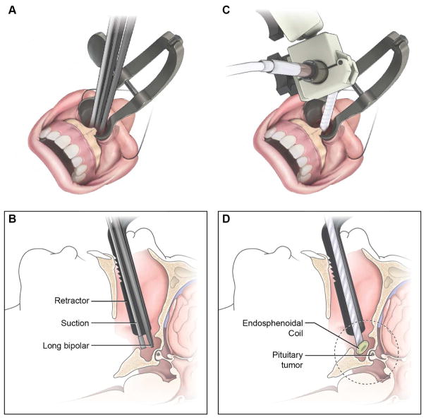
Figure 3.
Positioning of the ensdosphenoidal coil (ESC) within the surgical path created during TSS. Standard approach to the sella involves creating a surgical tract, removal of sphenoid wall and insertion of MRI self-retaining retractors (A). The surgical corridor allows for insertion of instruments such as suction and bipolar cautery devices (B). Following initial approach, the ESC can be placed within the surgical corridor (C) such that the distal coil reaches within the sphenoid sinus (D).

