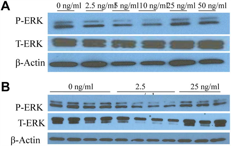
Figure 1. ERK activation in H-69 human cholangiocyte cells (A and B) by FGF19.

Western blotting of cholangiocyte phosphorylated (P-) and total (T-) ERK after a 24h incubation period with various concentrations of FGF19. Data represent pooled samples with n = 4 (A) and unpooled single samples (B).
