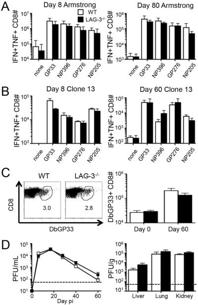
Figure 2. The complete absence of LAG-3 does not affect the number, function, or longevity of antiviral CD8+ T cell responses.
T cell responses and viral loads were measured in WT or LAG-3−/− mice following acute or chronic infection. (A) WT B6 and LAG-3−/− mice were infected with LCMV-Armstrong and the total number of splenic IFN-γ/TNF-α double positive CD8+ T cells following ex vivo stimulation with various LCMV peptides was measured by intracellular cytokine staining at days 8 (left) and 80 (right) post-infection (pi). (B–D) WT B6 and LAG-3−/− mice were infected with LCMV-Clone 13. (B) The total number of splenic IFN-γ/TNF-α double positive CD8+ T cells following ex vivo stimulation with various LCMV peptides was measured by ICS at days 8 (left) and 60 (right) pi. (C) Examples of Db/GP33 tetramer staining of CD8+ T cells in the spleens of either WT or LAG-3−/− mice at day 60 (left); the numbers indicate the percentage of tetramer+ cells among CD8+ T cells. The total number of splenic LCMV-specific CD8+Db/GP33+ cells in either WT or LAG-3−/− mice at day 60 pi (right). (D) The level of infectious virus was determined by plaque assay from serum samples over time (left) and liver, lung, and kidney tissues at day 60 (right). The dashed line represents the limit of detection. The data in panel A represent 9 mice from 3 independent experiments. All other panels represent 6 mice from 2 independent experiments.

