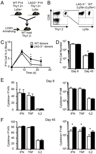
Figure 3. Direct LAG-3 signaling reduces the abundance of memory CD8+ T cells following acute viral infection.
A dual adoptive transfer approach was used to directly compare the effect of LAG-3 expression on CD8+ T cells following acute infection. (A) A mix containing 2×10³ Thy1.2+Ly5a+ WT P14 cells and 2×10³ Thy1.2+Ly5a− LAG-3−/− P14 cells mice was adoptively transferred into Thy1.1+ B6.PL mice; 4 days later the mice were infected with LCMV-Armstrong. (B) An example of the gating strategy used to identify the WT and LAG-3−/− P14 cells. After gating on CD8+Thy1.2+ cells, the transferred cells were distinguished by their expression of Ly5a. (C) The frequency of each population of P14 cells among blood leukocytes in mice that were bled repeatedly following infection. (D) The total number of splenic WT and LAG-3−/− P14 cells at days 8 and 45 pi. (E–F) The frequency (left) and total number (right) of cytokine+ splenic WT and LAG-3−/− P14 cells at day 8 (E) and day 40 (F) as measured by ICS after ex vivo stimulation with GP33-41 peptide. The day 8 data represent 5 mice from 2 independent experiments while the day 40 data represent 9 mice from 3 independent experiments.

