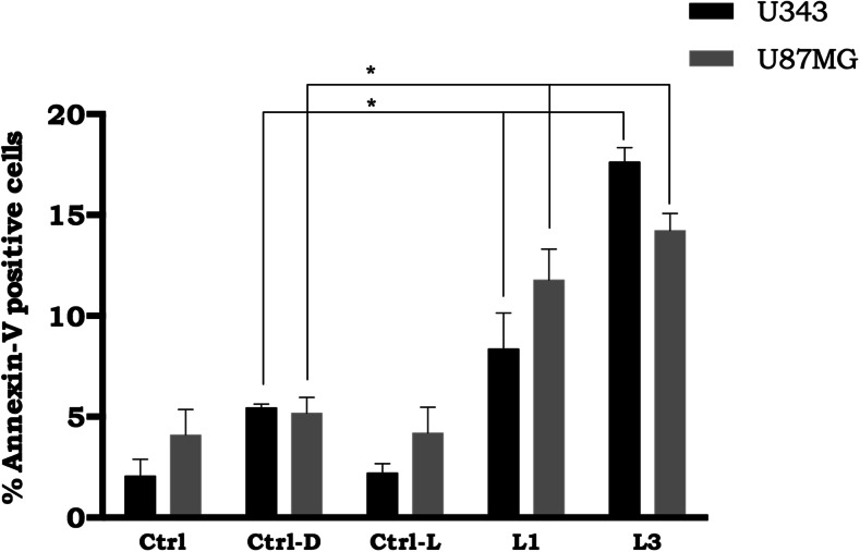
Fig. 6.
Cell death assay of glioblastoma lines by flow cytometry in the presence of NE/ClAlPc (0.5 μM) under different irradiation conditions after 48 h of treatment. Ctrl: Control samples cultured with 3% fetal bovine serum, Ctrl-D: control samples with NE/ClAlPc in the absence of sunlight light (darkness), Ctrl-L: control samples subjected to sunlight at a dose of 700 mJ/cm2 in the absence of NE/ClAlPc, L1: samples with NE/ClAlPc subjected to sunlight light at a dose of 100 mJ/cm2, L3: samples with NE/ClAlPc subjected to sunlight light at a dose of 700 mJ/cm2. Statistical significance was set at *p < 0.05

