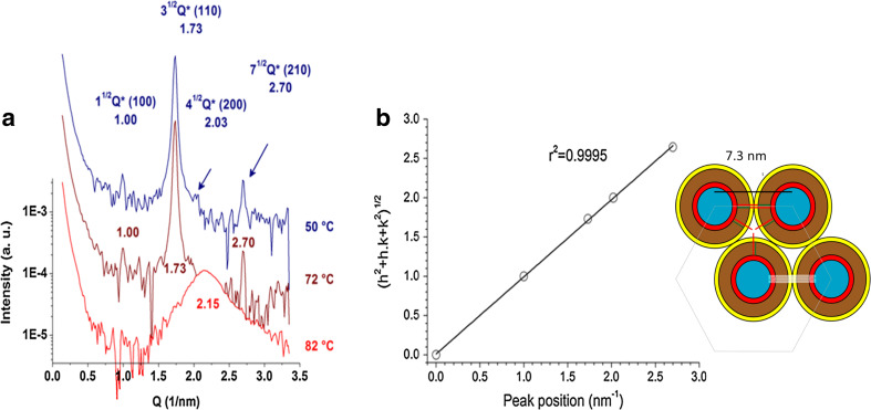
Fig. 6.
Structural analysis of 10:0 ceramide aqueous suspension. a SAXS patterns of 10:0 Cer at the indicated temperatures in the hexagonal phase (blue and brown) and disordered phase (red). Synchrotron radiation was used and spectra were acquired after 10 min of thermal stabilization. Measurements were carried out in triplicate with fresh samples. b Correlation of the position of the SAXS peaks with Miller indexes of a hexagonal crystal array. The scheme show a simplified model of the HII tubular micelles represented in cross-section. The main slabs are shown in color: blue for water, red ring for polar headgroup, brown ring for the non-interdigitated acyl chain region, yellow ring for the asymmetric (interdigitated) region of the acyl chains, green stick (acyl chain of 10 carbonyls), red stick for the acyl part of sphingosine and white is the packing frustration region. The remarkable feature of this cartoon is that the sphingosine methyl end practically fit close to the center of the space between the cylinders, relieving packing frustration. Adapted from Dupuy et al. (2017)

