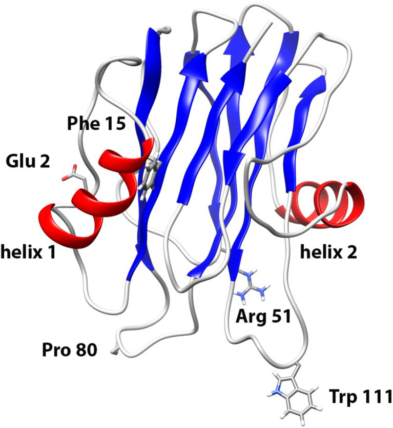
Fig. 1.
Structural elements of actinoporins. Ribbon representation of sticholysins I (StI) structure (Protein Data Base: 2KS4). α-Helices are represented by light gray ribbons, β-sheets are in dark gray and non-periodic structures are gray color. The amino acids Glu2, Phe15, Arg52, Pro80 and Trp111 replaced by Cys in the site-directed mutants are indicated in the figure. Images were produced with the UCSF Chimera program for Windows (Pettersen et al. 2004)

