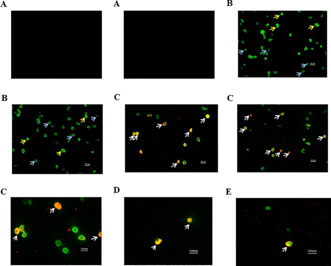
Fig 4. Representative pictures taken from fluorescence microscopy of isolated smooth muscle cells (positive for SM22α; green) and endothelin B receptor (positive ETB; red) from different tissue.
(A) IgG SM22α and IgG ETB (N = 3); (B) VSMC of pial vessels stained with SM22α, blue arrows-dim VSMC and yellow arrows-bright VSMC (N = 3); (C) VSMC of pial vessels stained with SM22α and ETB (N = 3); (D) VSMC of parenchymal vessels stained with SM22α and ETB (N = 3); (E) VSMC of retina vessels stained with SM22α and ETB (N = 3). The white arrows indicate cells positive for both SM22α (green) and ETB receptor (red).

