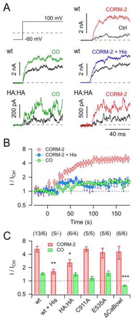
Fig. 1. Activation of KCa1.1 channels by CORM-2 and CO.
A) Current traces in response to the indicated pulse protocol recorded from inside-out membrane patches of HEK 293T cells expressing KCa1.1 wild type (wt) or mutant H365A:H394A (HA:HA) before (black) and 3 min after the application of either 50 µM CORM-2, 250 µM CO, or 50 µM CORM-2 in the presence of 1 mM histidine (colored). B) Time courses of the normalized steady-state current with 50 µM CORM-2 (without and with 1 mM histidine) or 250 µM CO application at time zero. Straight lines connect data points for clarity. C) Mean steady-state current increases by 50 µM CORM-2 (red) and 250 µM CO (green) for the indicated channel types and conditions. Data in B and C are means ± S.E.M. with n indicated in parentheses. Asterisks indicate difference to respective wild-type data: *P<0.05; **P<0.01; ***P<0.001.

