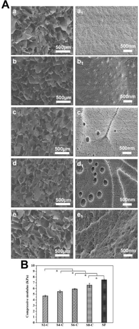
Figure 3.
Morphology of SF-CS scaffolds: (A) SEM images of scaffolds: (a–e) indicate the porous structures of SF, S2-C, S4-C, S6-C and S8-C samples, while a1-e1 show the nanotopography of the porous walls; (B) compressive modulus of the scaffolds in wet conditions. *Statistically significant P < 0.05.

