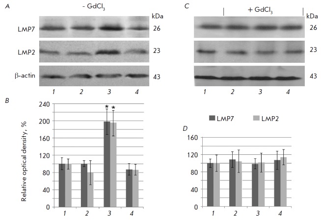
Fig. 1.
Content of the proteasome subunits LMP7 and LMP2 in clarified homogenates of false-operated rat liver on the 7th day after the introduction of a physiological solution (1) and in rat liver on the 1st day (2), 7th day (3), and 14th day after the beginning of DST induction (4) with preliminary injection of GdCl3 and without it. A, C – Western blots of subunits LMP7, LMP2 and β actin. B, D – Relative quantity (optical density of blots) of subunits LMP7 and LMP2 normalized to β actin content. The subunit quantity in the samples of false-operated animals was taken as 100%; means ± SEM are shown; significant difference at p < 0.05 and n = 5–6 in comparison with the false-operated control is indicated (*).

