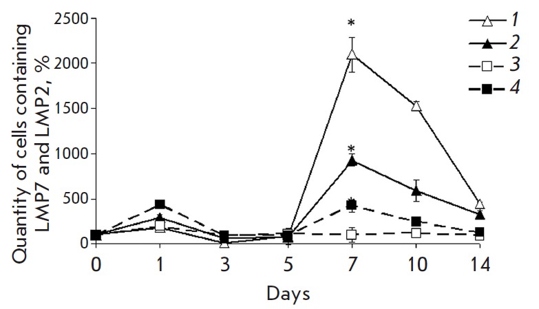
Fig. 3.

Change in the quantity of mononuclear cells containing the proteasome LMP2 subunit (filled symbols) and LMP7 subunit (empty symbols) in different time intervals after the beginning of DST induction in the liver of the 3rd (lines 1 and 2) and 4th rat groups (lines 3 and 4). On x axis, days after the beginning of DST induction are shown. The quantity of cells containing the LMP2 and LMP7 subunits in samples of the 1st group was taken as 100%. Significant difference at p < 0.05, n = 5 in comparison with basal level (group 1) (*).
