Figure 1.

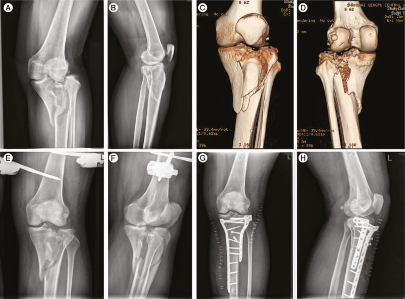
Case 1 undergoing external fixation and delayed open reduction and internal fixation. Preoperative X-ray imaging (A, B), 3D reconstruction imaging (C, D), postoperative CT scan (E–H).

Case 1 undergoing external fixation and delayed open reduction and internal fixation. Preoperative X-ray imaging (A, B), 3D reconstruction imaging (C, D), postoperative CT scan (E–H).