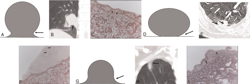
Figure 1.

Pictograms, CT images, and photopathologic features of various tumor borders. (A–C) Representative type 1, S or reverse S border with a blunt angle (arrow), in 80-y-old male with adenocarcinoma (arrows) invading of the elastic layer of the visceral pleura but without reaching the visceral pleural surface (PL1) (orcein stain, magnification ×40). (D–F) Type 2, sharp angle (arrow), in 54-y-old female with adenocarcinoma (arrows) invading the visceral pleural surface (PL2) (orcein stain, magnification ×40). (G–I) Type 3, concave border with a blunt angle (arrow), in 58-y-old male with adenocarcinoma (arrows) invading the elastic layer of the visceral pleura (PL1) (orcein stain, magnification ×40).
