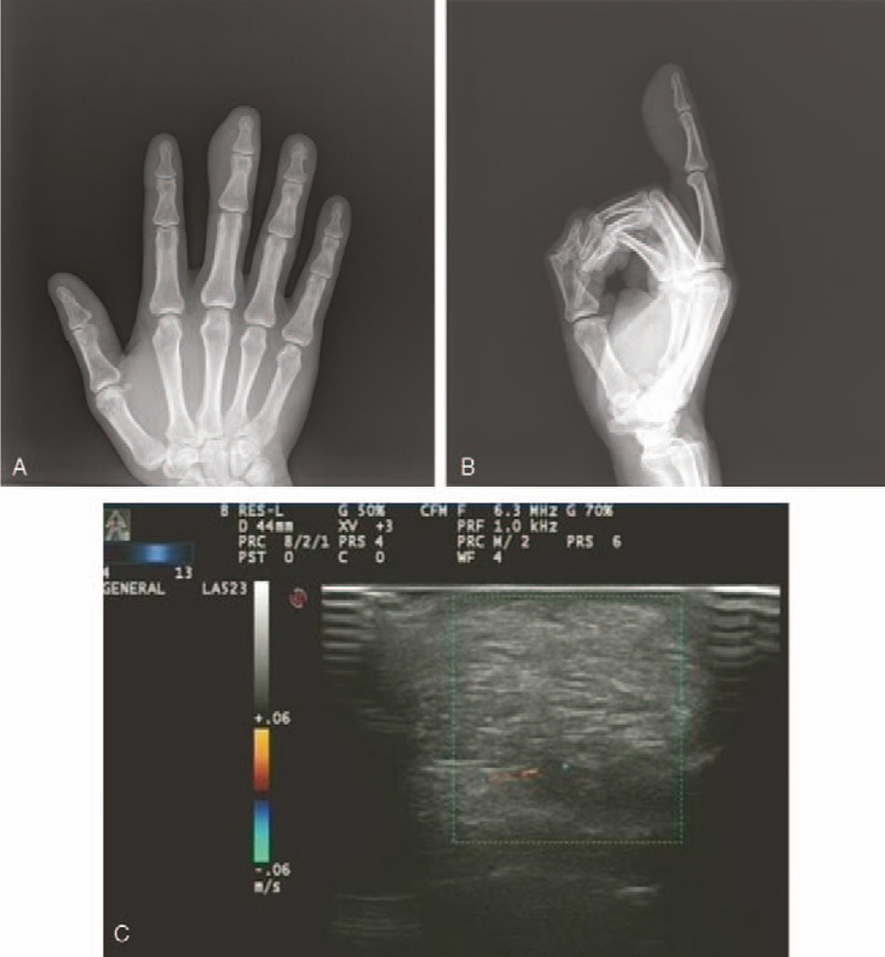
Figure 2.

A: Radiography of the middle finger shows a soft tissue swelling of low density over the distal phalanx (anteroposterior view). B: Radiography of the middle finger shows a soft tissue swelling of low density over the distal phalanx (lateral view). C: An ultrasound scan shows a well-defined hyperechoic subcutaneous mass and no internal flow is detected.
