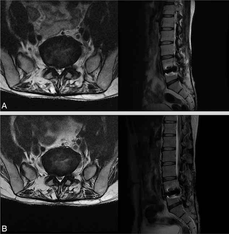
Figure 1.

(A) Axial (left) and sagittal (right) T2-weighted lumbar spine MRI 1 month after the onset of lower back and right buttock pain showed right central disc extrusion at L5–S1. The artifact observed in the L4–5 level is due to the metal implant used in the operation for herniated lumbar disc 10 years ago. (B) In the follow-up MRI performed 1 day after SELD, no newly developed lesions, such as hematoma, infection, and aggravated HLD, were observed. HLD = herniated lumbar disc, MRI = magnetic resonance imaging, SELD = trans-sacral epiduroscopic laser decompression.
