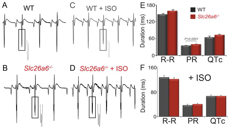
Figure 6. ECG recordings from age-matched WT and Slc26a6−/− mice reveal evidence of fragmented QRS complexes in the knockout mice.
A to D. Representative ECG traces. The insets showed the enlarged traces marked by the rectangular boxes demonstrating fragmented QRS complexes observed only in Slc26a6−/− mice. E. ECG parameters in control condition and with isoproterenol (ISO) (F).

