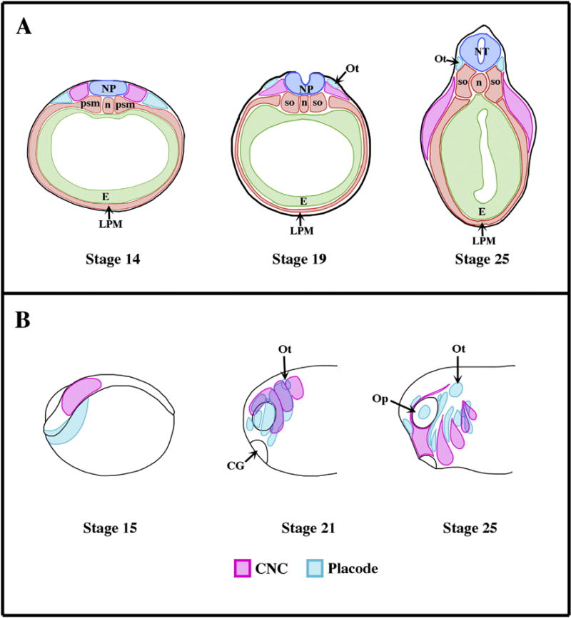
Fig. 1.

Origin and migration of the cranial neural crest cells in Xenopus laevis. (A) Schematics representing transverse sections though the prospective otic placode (Ot) of Xenopus embryos at the various stages of development (Nieuwkoop and Faber, 1967). Dorsal is up. The placodal ectoderm and neural crest prospective tissue emerge from the sensory layer of the ectoderm and are anatomically visible as soon as stage 14 (early neurulation). At stage 19 (late neurula stage) the cranial neural crest starts migrating ventrally (Sadaghiani and Thiebaud, 1987). (B) Schematic showing the relative positioning of CNC and the cranial placodes (Sadaghiani and Thiebaud, 1987; Schlosser, 2006). During the early neurula stage (stage 15) both placodal (blue) and CNC (pink) primordium appose one another. Starting with the early tailbud stage (stage 21), these territories are segmented into various subgroups that reflect their future differentiation. The placodal ectoderm secretes Sdf1, which attracts CNC. Once the CNC reaches a placode, CIL takes place and the placode migrates away (i.e. ventrally). Once the placodes and CNC are separated, the Sdf1-based chemoattraction reasserts itself. This “chase and run” contributes to the proper migration of the each of the CNC segments while the placodes lying in the path of the CNC (e.g. Epibranchial placodes) reach their proper dorsoventral location and are shaped into narrow strips of tissues. Ot: Placode; NT, neural tube; n: notochord; psm: presomitic mesoderm, so: somites; LPM: lateral plate mesoderm, CG: cement gland; Op: optic vesicle.
