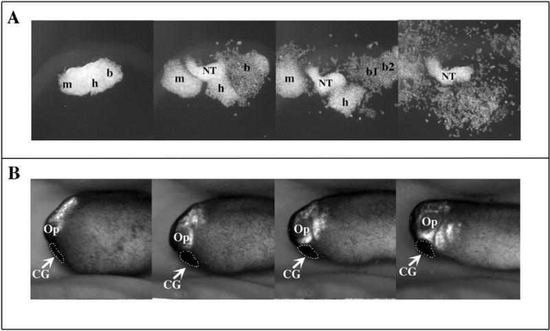
Fig. 2.

Migration of CNC cells in vitro and in vivo. GFP expressing CNC are explanted at stage 15 and either grown on a substrate of fibronectin in a saline media (A) or grafted into a wild type embryo (B) (anterior left, dorsal top). The migration of the CNC is followed by time-lapse photography (3 minute intervals, 10 h of total migration) and still pictures from these movies were extracted at regular time points throughout the movies. The migration of CNC in vitro (A) recapitulates the migration of the CNC observed in vivo (B) including the formation of segments (mandibular, hyoid, branchial), and the collective migration which is observed in the first 3 frames and the single cell migration which is observed in the last frame. Note that the explant in A was cut purposely large to demonstrate the formation of segments and therefore contains some neural tissue contaminant (NT). m: mandibular; h: hyoid segment b: branchial segments (b, b1, b2); CG: cement gland.
