Fig. 1.
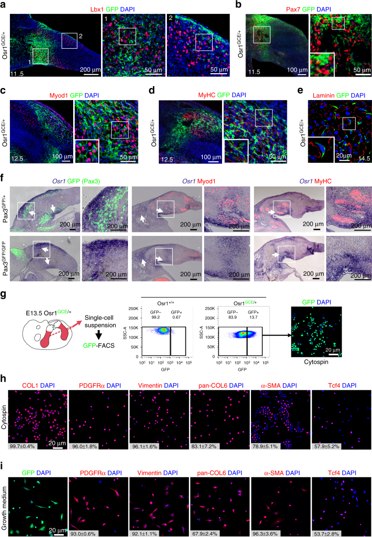
Osr1 is expressed in limb mesenchymal cells associated with myogenic cells and labels MCT cells. a–e Osr1 expression was assessed by immunolabelling for GFP on Osr1 GCE/+ embryos at the indicated stages. Osr1+ cells are found interstitial to Lbx1+, Pax7+ and MyoD1+ myogenic progenitors and interstitial to developing myofibres labelled either for Myosin heavy chain (MyHC) or Laminin. Note that in some developing muscles Osr1+ cells are abundant, while being scarce in others. Boxed regions (numbered in a) are shown in the panels to the right of overview images and are magnifications of the area. f Osr1 expression was analysed by in situ hybridization to sections of muscleless limbs of E11.5 (left), 12.5 (middle) and 13.5 (right) Pax3 mutant embryos. The overall expression pattern of Osr1 is similar in limbs of Pax3 GFP/+ and Pax3 GFP/GFP embryos. Arrows highlight strong Osr1 expression observed in control and muscleless limbs. Boxed regions are shown in the panels to the right of overview images and are magnifications of the area. g Schematic depiction of limb Osr1+ cell isolation procedure and examples of FACS plots. h Expression of fibroblastic markers in freshly FACS-isolated Osr1+ cells subjected to cytospin. (n = 3). i Osr1+ cells cultured in growth medium maintained the expression of fibroblastic markers (n = 3). Insets in h, i are percentage of COL1+, PDGFRα+, Vimentin+, Pan-COL6+, αSMA+ and Tcf4+ cells among the Osr1+ cell population. Values represent mean ± s.e.m. N-numbers indicate biological replicates, i.e. samples from different specimen

