Figure 7.
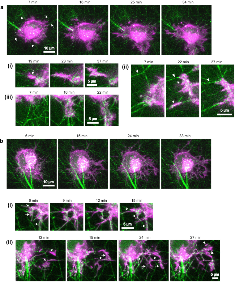
HT-1080 cell protrusions exhibit contact guidance along collagen I fibers under control or blebbistatin-treated conditions. HT-1080 cells, stained with the membrane dye DiI (magenta) were seeded in 1.2 mg/ml rat-tail collagen ECMs (green) and imaged over time. Times indicated in the figure are relative to the time of seeding. Primary panels (a and b) are maximum projections of z-stacks. The sub-panels (i,ii,iii) show selected frames and z-slices from regions of interest in the associate full-cell image. (a) Selected frames from Supplementary Movie 6, representative of seven independent experiments. (7–16 min) The cell initially protrudes in multiple directions. Stable protrusions coincide with centripetally aligned fibers (e.g. arrows). (16–25 min) Cell begins moving predominately in one direction (toward the bottom of the frame) and the associated protrusion/retraction activity compacts fibers and aligns them in the direction of migration (see A.iii and Supplementary Movie 6 for clearer depiction of fiber movement). (a.i) Protrusion progresses along an aligned fiber. (a.ii) Two protrusions progress along centripetally aligned fibers (arrows). The tensile forces applied during the protrusion/retraction cycles cause the movement and reduction in angle (alignment) of fibers (arrowheads). (a.iii) A protrusion causes fiber alignment and compaction. (b) Selected frames from Supplementary Movie 7 of a cell treated with 50 µM blebbistatin (representative of four independent experiments). The cell extends many thin and highly dynamic protrusions. Protrusions are not localized to areas of high matrix alignment, but a higher number of protrusions extend toward the bottom of the frame, possibly in response to the predominant alignment of the fibers. There is little cell-mediated fiber movement; instead, protrusions often follow fibers like tracks, even if they do not follow a straight line. (b.i) Protrusions progress along fibers at multiple angles to the cell. (b.ii) (12 min) Two protrusions appear (arrows), extending downward from a different z-plane (not depicted). (15–24 min) The two main protrusions split into multiple smaller protrusions (arrows) that progress along fibers at multiple angles to the cell. (27 min) A third main protrusion progresses along two, non-aligned fibers (arrowheads). Scale bars, 10 µm (main panels), 5 µm (sub-panels).

