Figure 5.
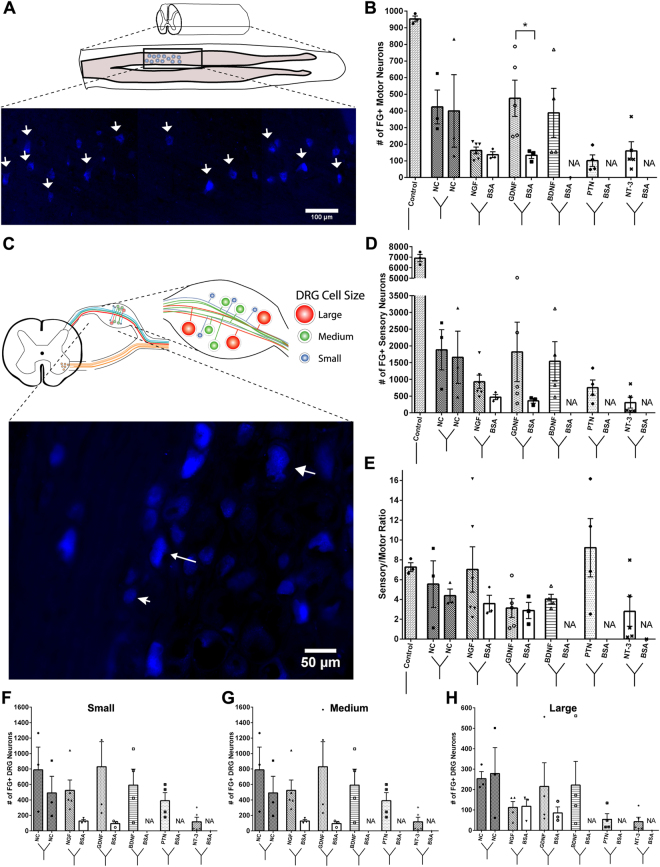
The effect of single guidance cues on the regeneration of motor and sensory neurons. (A) Schematic of the longitudinal section of the ventral spinal cord showing fluorogold positive cells (white arrows) (B) Quantification of FG+ cell in each of the split Y-nerve. GDNF had a significantly higher number of regenerated motor neurons compared to BSA. (C) Schematic of DRG soma size distribution and representative image of fluorogold positive cells of varying size. (D) Quantification of the total number of FG+ sensory neurons. (E) Single guidance cues did not significantly alter the sensory to motor ratio. (F–H) Distribution of small to large sensory neuron soma sizes showed no difference between the neurotrophic factor loaded microparticles and BSA loaded microparticles. Data represented as mean ± SEM. (*P ≤ 0.05). NA represents groups with failed retrograde tracing using FluoroRuby. (N = 3–6 animals/group).

