Figure 4.
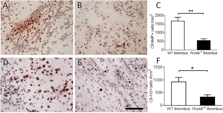
NET formation in thrombi 48 hours after IVC ligation was reduced in pcsk9 −/− mice compared to WT mice (n = 5 mice per group). (A,B) Representative photomicrograph of cathelicidin-related antimicrobial peptide (CRAMP) staining from WT (A) and pcsk9 −/− (B) mice. (C) Quantification of CRAMP-positive cells per unit area. (D,E) Representative photomicrograph of citrullinated histone H3 (Cit-H3) staining from WT (D) and pcsk9 −/− (E) mice. (F) Quantification of Cit-H3-positive cells per unit area. *P < 0.05. **P < 0.01. Scale: 50 μm.

