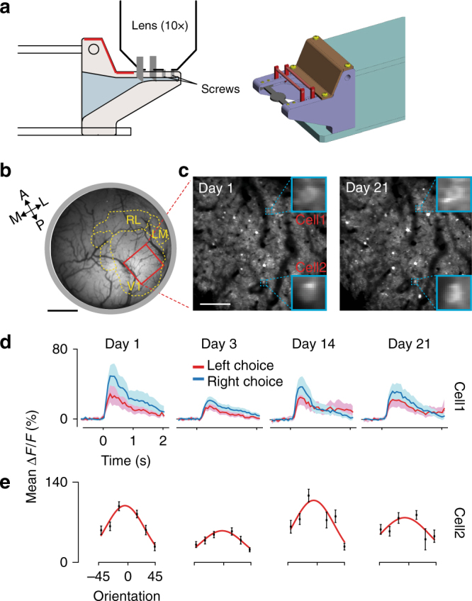
Fig. 5.

Compatibility of the setup with two-photon imaging. a Left panel: side view schematic of the latching unit for physiology with a ×10 objective lens for two-photon imaging. The two vertical gray segments indicate the latching pins (Supplementary Fig. 6). Right panel: 3D rendering of the latching part of the unit. b Wide-field GCaMP8 fluorescence image at the cortical surface. Red rectangle, ROI within V1. Scale bar 1 mm, dotted yellow lines and labels are segmented visual areas (Methods). c Repeated imaging of the same ROI across different days. Blue squares are magnified views of two example cells. Scale bar, 200 μm. d Visually evoked responses during the behavioral task recorded from cell1 shown in c. ΔF/F trial averages for left/right choices (red/blue lines). Shaded areas, 95% confidence interval. Time-out trials were not included in the data. e Responses to oriented gratings (30° diameter) from cell-2 shown in c. Error bars are s.e.m. Red curves, Gaussian fit to the data. Original technical drawings in a edited with permission from O’ Hara & Co., Ltd.
