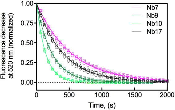
Figure 3.

Determination of the rate of spontaneous nanobody dissociation from BtuFfluo (k off) at pH 7.5 and 23 °C. Illustrated are the kinetics of displacement of nanobodies from BtuFfluo by Cbl. Shown is the normalized fluorescence decrease at 520 nm over time (raw data is shown in Supplementary Figure 3). BtuF (10 nM) was incubated with 10 nM nanobody prior to the addition of excess Cbl. The shown fluorescence traces correspond to nanobody displacement by Cbl. Identical rate constants were obtained at three different Cbl concentrations ((○) 1 μM, (∆) 5 μM and (□) 10 μM), showing that the fluorescence decrease directly reported nanobody dissociation from Cbl. The obtained k off values are listed in Table 1. We also observed a rapid fluorescence decrease during the dead time of manual mixing, presumably caused by the binding of Cbl to free BtuFfluo. This rapid phase is not shown for clarity.
