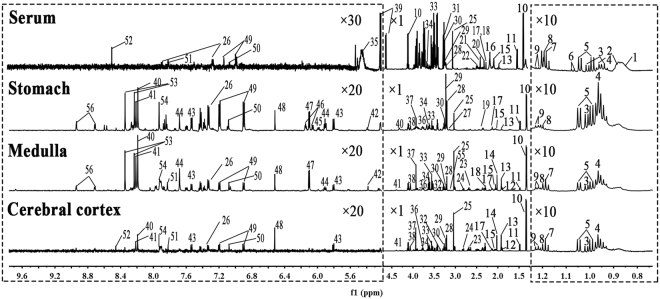
Figure 3.
Typical 1H NMR spectra of serum and aqueous extracts from stomach, medulla and cerebral cortex tissues. (1, Low density lipoprotein; 2, Very low density lipoprotein; 3, Isoleucine; 4, Leucine; 5, Valine; 6, 2-Ketobutyric acid; 7, Ethanol; 8, β-OH-butyrate; 9, Methylmalonate; 10, Lactate; 11, Alanine; 12, γ-Aminobutyrate; 13, Acetate; 14, N-acetyl aspartate;15, Glutamate; 16, O-acetyl glycoprotein; 17, Glutamine; 18, Methionine; 19, Glutathione; 20, Acetone; 21, Acetoacetate; 22, Citrate; 23, Aspartate; 24, N,N-dimethylglycine; 25, Creatinine; 26, Phenylalanine; 27, Ethanolamine; 28, Choline; 29, Phosphocholine; 30, Glycerophosphocholine; 31, Betaine; 32, Inositol; 33, Glycine; 34, Glycerol; 35, Glycogen; 36, Serine; 37, Phosphocreatine; 38, Adenosine monophosphate; 39, Glucose; 40, Hypoxanthine; 41, Inosine; 42, Allantoin; 43, Uracil; 44, Uridine; 45, Uridine diphosphate glucose; 46, NADP+; 47, Inosinic acid; 48, Fumarate; 49, Tyrosine; 50, Histidine; 51, Methylhistidine; 52, Formate; 53, Adenosine; 54, Xanthine; 55, Malonic acid; 56, Nicotinamide).

